Why Dentists Use a Microscope for Root Canal Treatment
What a dental operating microscope lets a dentist see during root canal treatment: canal anatomy, cracks and calcified passages.
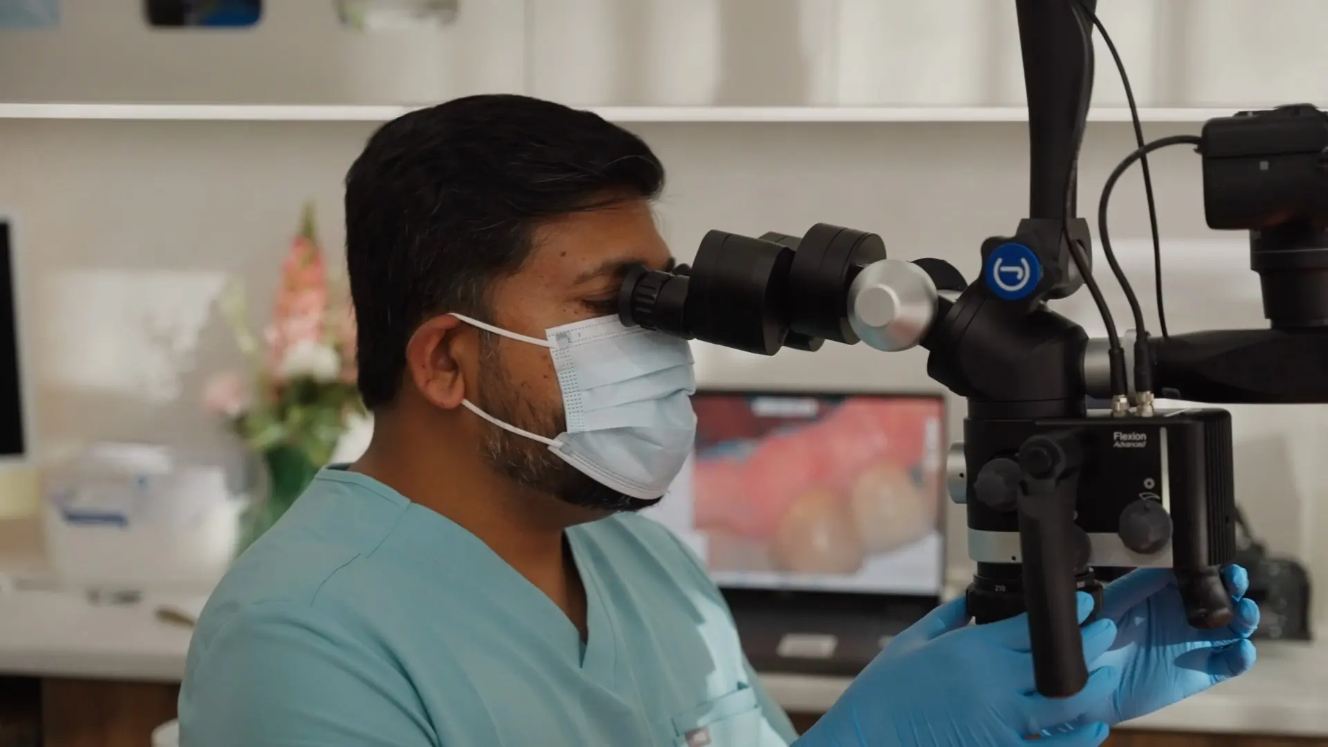
The inside of a tooth is smaller than most people imagine. The canals that hold the nerve and blood supply of a molar can be narrower than a human hair, they curve, they branch, and they are not always where the textbook diagram suggests. Root canal treatment involves cleaning those passages thoroughly, which raises an obvious question: how does anyone see well enough to do that?
Part of the answer is a dental operating microscope. It is a piece of equipment that gets talked about surprisingly little outside the profession, partly because it is undramatic and partly because it is easier to sell a story about comfort than about optics. This article explains what a microscope actually is in a dental context, what it lets a dentist see, and where it fits into root canal treatment. It is general information only and is not a substitute for a consultation with a qualified practitioner.
A microscope is not the same as loupes
Most dentists work with magnifying loupes (the small telescopic lenses mounted on a pair of glasses), usually paired with a headlight. Loupes are genuinely useful and are standard equipment across modern dentistry.
A dental operating microscope is a different instrument. It is a large, counterbalanced unit mounted on a stand or arm, positioned over the treatment area, with a much greater range of magnification than loupes provide and light delivered straight down the same optical path the dentist is looking through. That last detail matters more than it sounds: because the light travels coaxially with the line of sight, it reaches into a narrow opening without the shadow that an angled headlight casts. Looking into a root canal is a bit like looking down a well. An angled torch lights the wall; a light coming from directly behind your eye lights the bottom.
At Dental Embassy in Braddon, Dr Srikanth Karumuri works with a dental operating microscope. Its use is case-selected rather than automatic, though in practice most root canal treatment is examined microscopically.
What magnification lets a dentist see
The practical value of a microscope is not that it makes the tooth look bigger. It is that certain features are difficult to identify reliably without it. Under magnification and coaxial light, a dentist is better placed to identify:
- Canal openings that are hard to find. Molars commonly have three or four canals, but an extra canal is not unusual: a second canal in the mesiobuccal root of an upper molar is a well-recognised example. A missed canal is one of the more common reasons a treated tooth stays symptomatic.
- Isthmuses and connecting passages. Canals are not always neat separate tubes; they can be joined by narrow ribbons of tissue that need cleaning too.
- Calcified canals. In older teeth, or teeth that have been through years of low-grade irritation, canals can narrow until they are extremely difficult to locate.
- Cracks in the tooth structure. A fine crack running down a wall can change the whole treatment discussion, and it is much easier to see under magnification than with the naked eye.
- The condition of previous work. In a tooth that has been treated before, magnification helps in assessing what was done and what is still there.
None of this is a promise about how any individual tooth will respond to treatment. Teeth vary enormously, and so do the situations they present in. What magnification changes is the amount of information available while the work is being done.
How it changes the appointment itself
Working under a microscope alters the ergonomics of treatment. The dentist sits upright, looking through the optics rather than leaning over the patient, which is one reason microscope work is often described as slower and more deliberate. For you as the patient, the visible differences are modest: the equipment sits above and behind you, you may be asked to stay quite still while the dentist is working at high magnification, and appointments may be planned with more time allowed.
That deliberate pace is worth knowing about in advance, because “careful and unhurried” and “long appointment” describe the same thing from different angles. If sitting still for an extended period is the part you find difficult (and for a lot of people it genuinely is), that is worth raising when treatment is planned. Options for managing longer appointments are covered in our guide to getting treatment done in fewer visits with sedation.
Where it matters most
Magnification is useful across a lot of dentistry, but the case for it is strongest where the working space is smallest and least visible. Root canal treatment is the clearest example, which is why microscopes became established in endodontics before they spread to general practice.
It also has a role in assessing a tooth that has had root canal treatment before and has become symptomatic again. Those cases turn on finding something that was not addressed the first time (an untreated canal, a crack, a blockage), and that is a task where seeing more is the whole point. We cover this situation in more detail in our article on root canal retreatment.
What to ask about it
If you are weighing up root canal treatment, the microscope is not really the thing to make a decision on by itself. More useful questions are practical ones: what has been found in your particular tooth, what the options are, how many visits are likely, and what happens if the tooth turns out to be less saveable than hoped. Our article on how the root canal or extraction decision gets made walks through that reasoning.
People in the inner north tend to arrive at these conversations from two directions. Braddon, Turner and Civic are heavily rented (around 60 per cent of Braddon households and 62 per cent in Civic, on the 2021 Census), with median ages of 30, 31 and 27, and renters who have moved cities often turn up with a tooth that has been grumbling for a while and no local dentist to call. From the older, more settled suburbs like Ainslie, where the median age is 42, the more common story is a tooth that was filled decades ago and has finally given up. Both are ordinary situations, and neither needs an apology.
Root canal treatment at Dental Embassy
Dental Embassy is on Northbourne Avenue in Braddon, a short walk or light rail trip from Civic and the inner-north suburbs. If you have been told you need root canal treatment, or you have a tooth that has been aching and you would like to understand your options, the starting point is an assessment of that specific tooth.
You can read more on our root canal treatment page, call us on 1300 000 230, or get in touch to arrange a consultation.
Have a question about your own situation?
Book a consultation with the team at Dental Embassy in Braddon. We’ll assess your teeth, explain your options, and answer your questions before anything is decided.