What a Wisdom Tooth X-Ray Actually Shows, and Why It Decides Everything
What a dentist reads on a wisdom tooth x-ray: angle, depth, root shape and nerve position, and when a flat image is not enough to plan a removal safely.
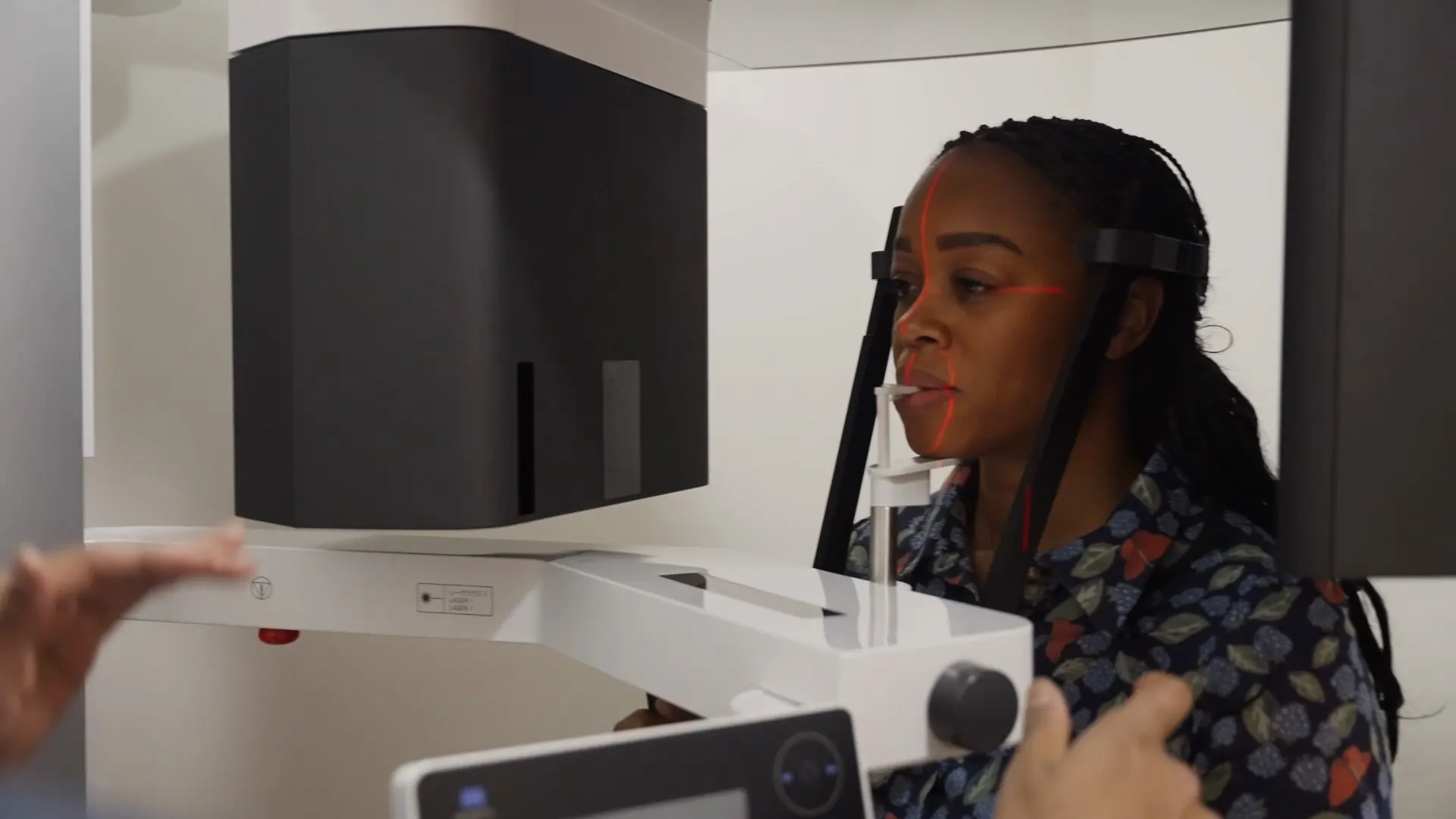
Most conversations about wisdom teeth start with a symptom and end with an x-ray. That order is worth noticing, because the symptom tells you something is wrong and the image tells you what to do about it.
If you have been shown a wisdom tooth x-ray and nodded along without really seeing what your dentist was pointing at, this explains what is being read. It is general information rather than a substitute for having your own images assessed.
The first image: an OPG
An OPG, sometimes called a panoramic x-ray, captures both jaws and all the teeth in one wide image. For wisdom teeth it is the standard starting point, because it shows all four at once along with the surrounding bone.
From that single image a dentist can establish four things.
The angle. Whether the tooth is upright, leaning forward into its neighbour, leaning backwards, or lying on its side. Each pattern carries a different risk of causing trouble and a different level of surgical difficulty.
The depth. Whether the tooth has broken through the gum, is partly covered, or is fully enclosed in bone. This matters more than most people expect, and it is covered below.
The root shape. Straight, curved, splayed or fused roots all behave differently during removal. Curved or splayed roots need a more careful approach because they resist coming out in one direction.
The neighbour. Whether the second molar in front is being damaged. Decay on the back surface of that tooth, in a spot no toothbrush reaches, is one of the strongest arguments for removing a wisdom tooth, and it is frequently invisible without an x-ray.
Why depth changes the whole picture
A fully buried wisdom tooth is sealed away from the mouth. Bacteria cannot reach it, and it will often sit quietly for decades causing nothing.
A partly erupted one is a different situation entirely. Where a flap of gum covers part of the crown, it creates a pocket that cannot be cleaned. Bacteria collect there, the gum becomes inflamed, and the result is pericoronitis: swelling and soreness at the back of the jaw, often with a bad taste and difficulty opening.
It typically settles with rinsing or antibiotics, then returns, because the pocket that caused it has not changed. That recurring cycle is one of the clearest indications for removal, and it is visible on the image before it has happened to you even once.
When a flat image is not enough
Lower wisdom teeth carry a question an OPG cannot always answer, and this is where the imaging becomes genuinely decisive.
The inferior alveolar nerve runs through the lower jaw and supplies sensation to your lip and chin. On a flat x-ray, a wisdom tooth root and that nerve canal can appear to overlap simply because they are at different depths and the image has flattened them into one plane. Overlapping on the picture does not necessarily mean touching in the jaw.
Where that overlap appears, we take a CBCT scan, which is a three-dimensional image. It shows whether the roots genuinely wrap around the nerve or merely sit near it, and that distinction changes the plan. It may change the surgical approach, and occasionally it is the reason we recommend leaving a tooth where it is, because the risk of removal outweighs the risk of keeping it.
Being told which group you are in is reasonable to expect, and we go through the images with you rather than summarising them.
What the x-ray can also show
A few less obvious findings turn up on wisdom tooth imaging.
Cyst formation. A widening dark area around the crown of a buried tooth can indicate a cyst developing in the follicle. These are usually painless and can quietly destroy bone, which is a strong reason for removal and a good argument for imaging even when nothing hurts.
Bone loss behind the second molar. A wisdom tooth pressing against its neighbour can cause bone loss on that side of the second molar, threatening a tooth far more valuable than the one causing the problem.
Resorption. Occasionally a wisdom tooth erodes the root of the tooth in front. This is silent and only visible on an image.
Nothing at all. Worth saying, because it is a common and legitimate outcome. Plenty of impacted wisdom teeth show no sign of any of the above, and monitoring them at regular check-ups is a perfectly defensible plan.
Why the image argues for a decision either way
The useful thing about basing this on imaging rather than symptoms is that it cuts in both directions.
It removes the case for taking out four wisdom teeth as a routine, because if the x-ray shows no problem developing, there is no argument to act on. It also removes the case for waiting until something hurts, because several of the findings above cause damage without producing symptoms until late.
There is one honest caveat about timing. When removal is genuinely needed, it is usually more straightforward at a younger age, because roots are shorter and bone is less dense. That is biology rather than a sales argument, and it is a reason to keep watching rather than a reason to operate now.
Around here
A large share of the wisdom tooth assessments we do are for people in their late teens and twenties, which is simply when wisdom teeth declare themselves. The ANU campus at Acton has a median age of 19, and Braddon and the city sit at 30 and 27, so that age band is heavily represented in our immediate catchment.
If your images do suggest removal, timing it around a semester break or a quieter work period is worth planning for, since recovery from a surgical extraction usually means two to three days of taking it easy.
What to ask
Ask to see the image, and ask which tooth is which. Ask what the angle is and whether the tooth in front is affected. For lower teeth, ask whether the roots are close to the nerve and whether a CBCT is warranted. And ask directly whether monitoring is a reasonable option in your case, because sometimes it is.
More detail on the condition sits on our impacted wisdom teeth page, the procedure and recovery on wisdom teeth removal, and the sedation options on sedation dentistry.
Have a question about your own situation?
Book a consultation with the team at Dental Embassy in Braddon. We’ll assess your teeth, explain your options, and answer your questions before anything is decided.